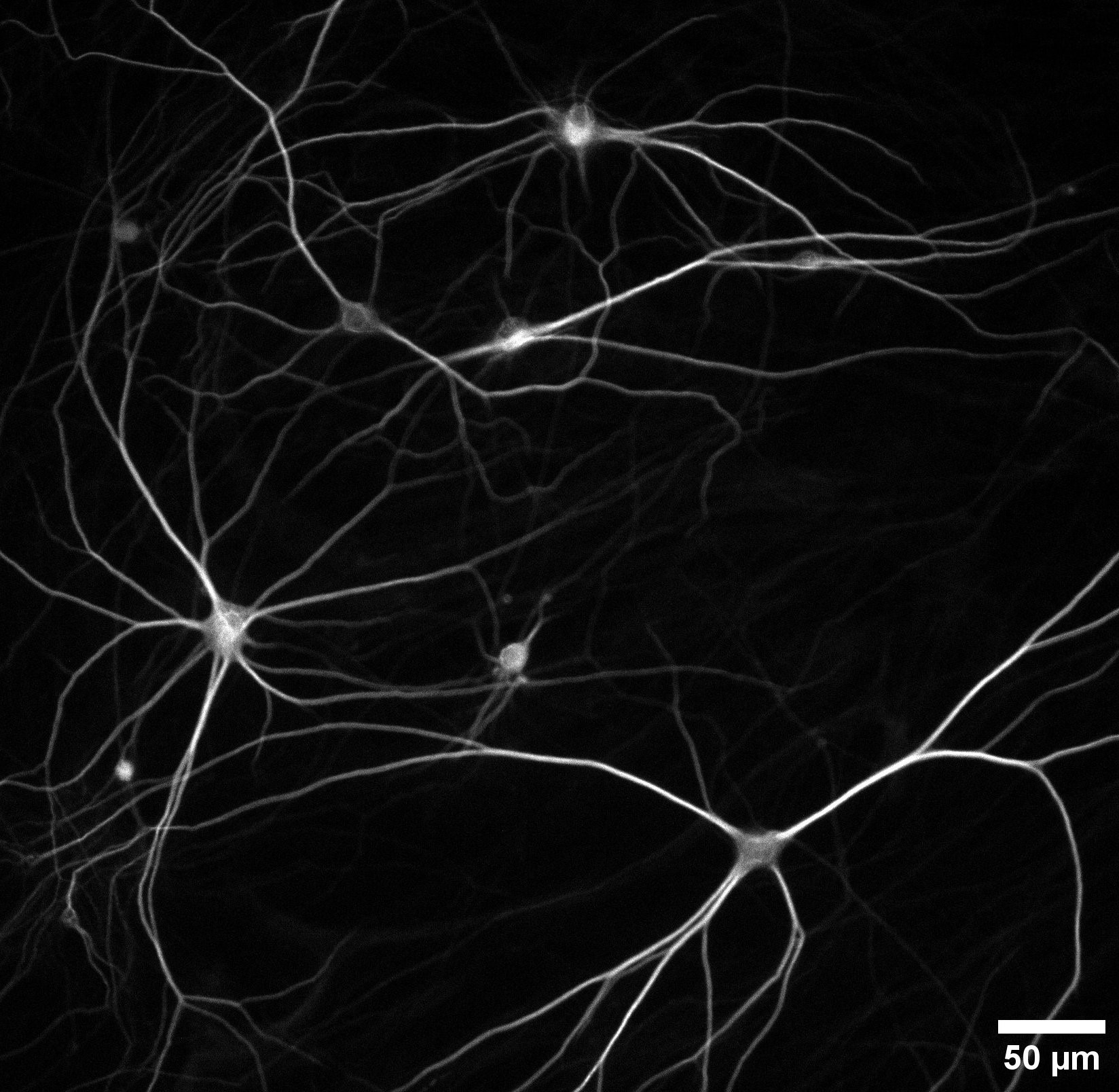
In this edition of Beauty of the Brain, you can see human neurons that have been grown in the laboratory from skin cells of Parkinson’s patients. It almost looks like a work of art, but these are real human brain cells that have been grown in the laboratory. Researchers at the Donders Institute do not create these neurons directly from brain tissue, but start with skin cells from healthy people as well as Parkinson’s patients. These skin cells are reprogrammed into a stem cell, which can then develop into all kinds of cells, including neurons.
In this case, the cells have been genetically programmed to become glutamatergic neurons, an important type of brain cell that transmits signals. The image mainly shows the dendrites, branched extensions that connect neurons to each other. They form a kind of network of wires through which information can flow.
The cells in this image are already about sixty days old. That may sound short, but in the laboratory that is quite a long time: the neurons are then “mature” enough to form networks, create synapses and communicate with each other. This makes them very similar to the cells in our own brains.
These cultured neurons are particularly special because they originate in part from people with Parkinson’s disease. The Kuijpers lab uses them to investigate what goes wrong in the early stages of the disease. This is because Parkinson’s is usually only discovered once brain cells have already died. By examining before the major damage is visible, researchers hope to better understand how the disease starts and ultimately discover ways to slow down or even prevent its development.
This specific image was created by postdoctoral researcher Eline van Hugte. She was able to make the cells visible with a simple stain and beautifully capture their typical branched shape. The result shows that science not only yields knowledge, but also images that reveal the beauty and fragility of our brain.
More Beauty of the Brain